Braincase Anatomy
Homepage > Dinosaur Anatomy -
Braincase Anatomy
The braincase of the dinosaurs is generally one of the most poorly understood regions of the dinosaur skeleton. Many of its components are fragile and easily destroyed, some of which do no ossify. Advances in science have lead to the usage of non-invasive computerized tomography (CT) scanning. This method gives researchers a clear sectional image without the risks of damaging the specimen.
The braincase houses the brain; by studying its anatomy one can get the overall size of the animals brain, to help calculate the animals intelligence. Through the delicate bone components, many process and bone openings can be seen, helping the researchers understand or get an idea of the animals sensory abilities.
•The braincase is usually fused up completely in mature animals, and most of its sutures are difficult to see.
•Dermal bones include the frontals and parietals on the roof of the skull. The frontal bones have usually well defined impressions of the olfactory tract and cerebral hemispheres.
•The occiput is normally formed by the supraoccipital, the basioccipital, and a pair of exoccipitals, these bones form the margin of the foramen magnum. The supraoccipital makes the dorsal margin of the foramen magnum, the sides are formed by the paired exoccipitals, and a small contribution of the ventral margin is composed by the basioccipital.
•A prominent knob-like process named the occipital condyle, is a rounded protuberance forming the ball of a ball and socket joint by which the head rotates on the neck, it articulates with the first cranial vertebra, the "atlas".
•The floor of the braincase ossifies with the basal plate of the chondrocranium into the basioccipital posteriorly and the basisphenoid anteriorly.
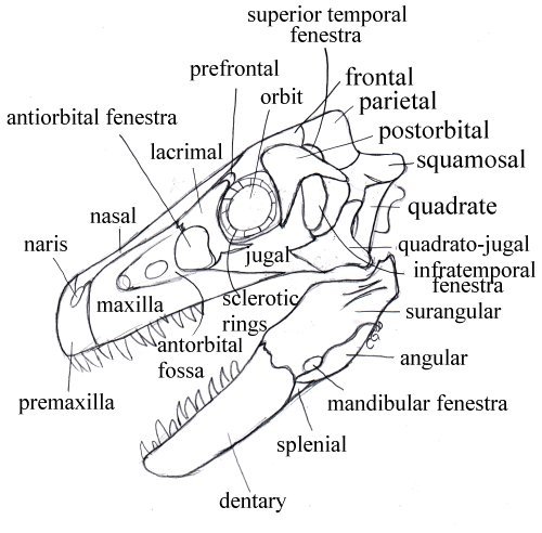
Dromaeosaurus albertensis skull with labeled parts.
Some dinosaurs had special structures inside of the skull. These dinosaurs were called Lambeosaurines, or crested hadrosaur dinosaurs. The largest crested of the lambeosaurines, Parasaurolophus, had a complex highway of tubes inside of its head, probably used for resonating sounds. These sounds may have been used for mating rituals, warnings to other members of the herd, or to help the Parasaurolophus recognize each other.
Braincase Bones
The supraoccipital is essentially an unpaired dermal bone of the occiput.
It is a bone found only in tetrapods and develops by ossification of a
membrane joining the two otic capsules. The supraoccipital contacts the
parietals of the skull table dorsally and the exoccipitals ventrally.
It usually forms at least the dorsal edge of the foramen magnum.
The exoccipitals are paired bones of the occiput. They derive from the neural arch elements of embryonic vertebrae which have been incorporated into the braincase. Dorsally, the exoccipitals contact the supraoccipital and the foramen magnum. Ventrally, they contact the basioccipital. The exoccipitals often form part of the occipital condyle.
The opisthotic is the posterior of the two bones making up the otic capsule and is usually fused to the prootic, the anterior otic bone. The opisthotic is an endochondral bone. It is usually the largest contributor to the paroccipital process and to the structures of the middle and inner ear. It normally contacts all of the occipital bones and the prootic, and may contact the basisphenoid. It often forms part of the edge of the foramen ovale, the door to the inner ear.
The paroccipital process may be considered a process of the opisthotic, but the process may be formed by bones in addition to, or even instead of, the opisthotic. The paroccipital process runs horizontally (and sometimes dorsally) across the back of the skull. It joins the occiput and braincase to the quadrate, squamosal and the other dermal bones of the "cheek."
The foramen ovale ( fenestra ovalis) is the only one of the various holes in Bob's head which makes our list of basic bits and pieces. It is a membrane covered manhole between the middle ear and inner ear. See, generally, The Ear. It is covered by the footplate of the stapes. The foramen ovale is normally located anterodorsal to the paroccipital process, between the prootic and opisthotic.
The stapes is an incarnation of the hyomandibular. Originally, it may have been the main upper element of a gill arch. It later appears as the hyomandibular, an accessory jaw element. In early tetrapods, it becomes a stout bone bracing the braincase against the quadrate. As the paroccipital process took over this function, the stapes was reduced, eventually becoming specialized for hearing as the columella, in sensible amniotes, or the stapes, in mammaliforms. The stapes bears a footplate which fits over the foramen ovale.
The occipital condyle is a rounded protuberance forming the ball of a ball and socket joint by which the head rotates on the neck. It is usually formed by some combination of the basioccipital and the exoccipitals.
The basioccipital is an unpaired median bone of the occiput which derives from the centra of embryonic vertebrae which have been incorporated into the braincase. It forms the floor of the braincase under the posterior part of the otic capsule. It contacts the exoccipitals dorsally and almost always forms at least part of the occipital condyle.
The basioccipital tubera are a pair of ventrolaterally directed blobs descending from the basioccipital. They are sometimes simply referred to as "basal tubera." However, the basisphenoid may also bear tubera. Presumably the basioccipital tubera act as attachment sites for ligaments stabilizing the head on the neck.
The basipterygoid processes are (despite the name) processes of the basisphenoid. They act to join the braincase to the palate. In many basal tetrapods and their ancestors, this was a moveable articulation. In most derived tetrapods, it simply staples the braincase to the palatal bones.
The pterygoid is not, of course, a braincase bone. It is the dermal palatal bone which grew up over the old palatoquadrate and eventually took over many of its functions. It is a very complex and interesting bone which, fortunately, we can skip over for present purposes. We have more than enough complex and interesting stuff to go 'round just dealing with the braincase.
The basisphenoid forms the floor of the braincase anterior to the basioccipital. Ventrally it is covered by a dermal bone, the parasphenoid. The fusion between these two is so close that some workers refer to the complex as the "parabasisphenoid." The basisphenoid gives rise to the basipterygoid process and other structures dealt with elsewhere.
The parasphenoid is the dermal bone normally found fused to the basisphenoid on its ventral surface. The parasphenoid generally extends far anteriorly on the midline of the palate as a narrow cultriform process. The upper surface of the parasphenoid (and the vomers) may be associated with the olfactory tracts. However, the anterior braincase is normally unossified in tetrapods.
The epipterygoid is another misnomer. Like the quadrates, the epipterygoids are ossified portions of the palatoquadrate (the original upper jaw which, like the hyomandibular, is homologous with an upper gill arch segment). They often appear to arise from the pterygoid, but do not. The epipterygoids are the true, old stuff of the palatoquadrate, while the pterygoid is but common dermal bone with pretensions. In fact, the epipterygoids are the original braincase articulations of the palatoquadrate. They demonstrate this ancient nobility by rising up in a graceful curve to reach the bones of the skull roof, like the last remaining columns in the abandoned temple of some forgotten god.
The prootic is the anterior of the two endochondral bones making up the otic capsule. It is usually fused to the other otic capsule component, the opisthotic. The extent and geometry of the prootic are quite variable. In addition to its fusion with the opisthotic, the prootic may contact the basisphenoid and any of the elements of the occiput.